- Empty cart.
- Continue Shopping


AccuTrack™
Our hardware-based eye tracker, compensates for blinks, loss of fixation and involuntary eye movements during scans reducing artifacts.

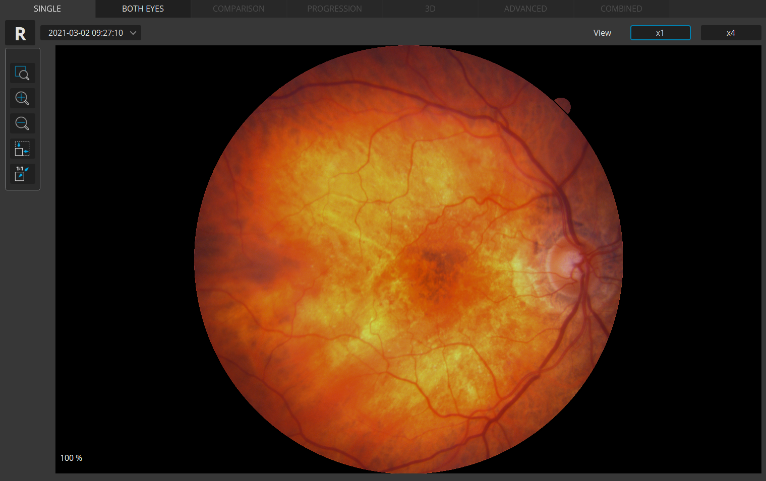
Auto Functions
Simplifying operation with the push of a button to auto-postion, auto-align, auto-focus, and auto-capture.
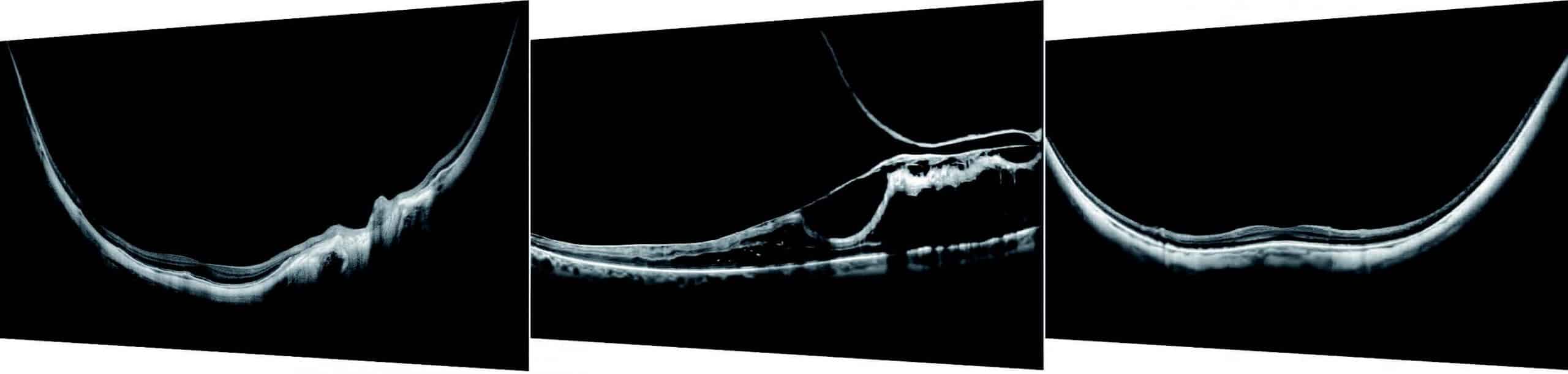
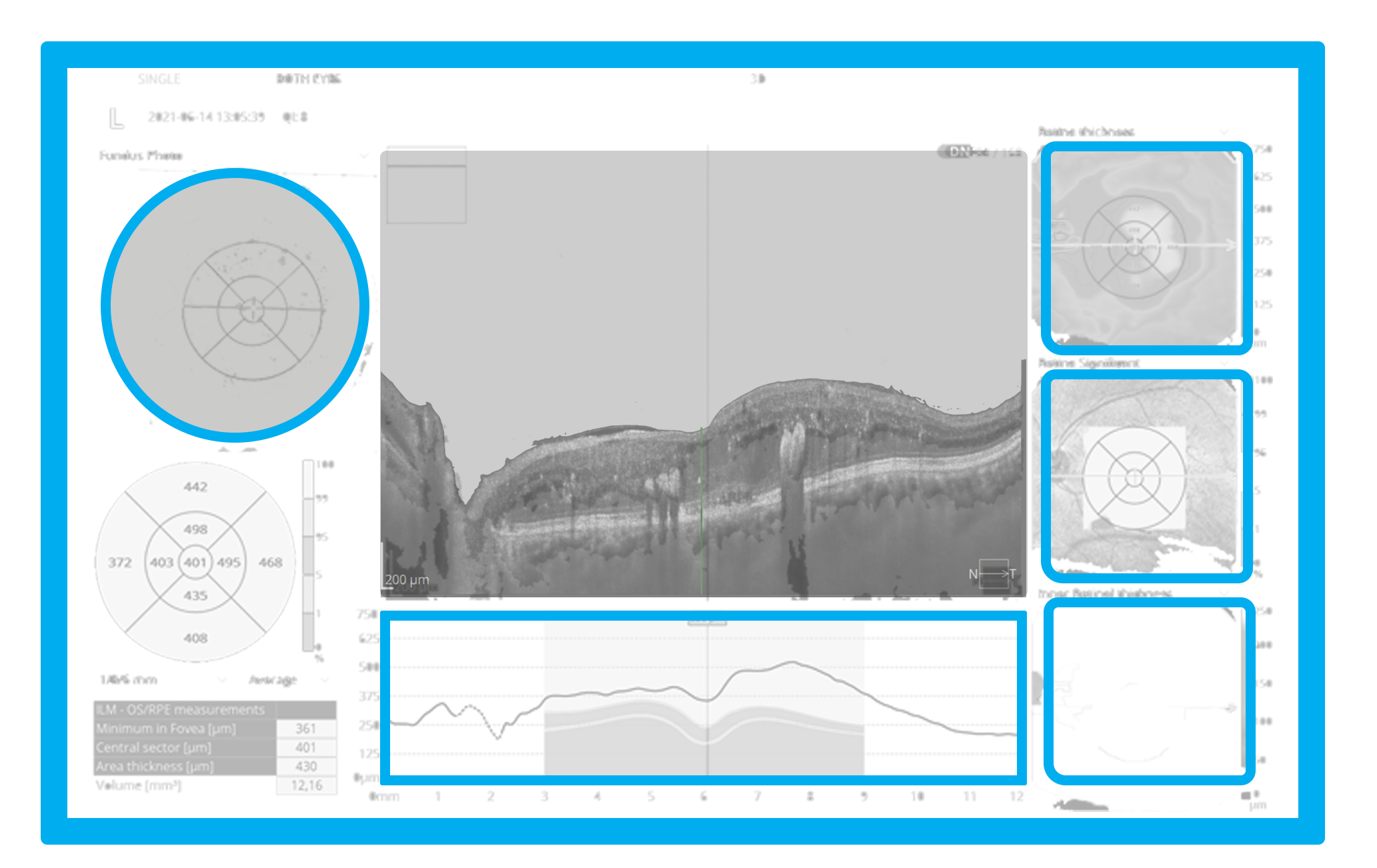
A.I. DeNoise
An advanced artificial intelligence (A.I.) algorithm removes noise from the tomogram for the highest image quality.
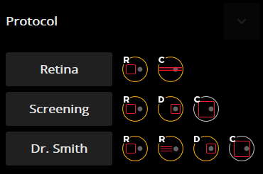
Custom Scan Protocols
Save time and never miss a scan. Create a custom preset group of scans and let the REVO capture all scans in order.
Motion Correction
The software-based motion correction (MC) compensates for involuntary eye movements and blinks by capturing two scans and generating a motion corrected scan when necessary.
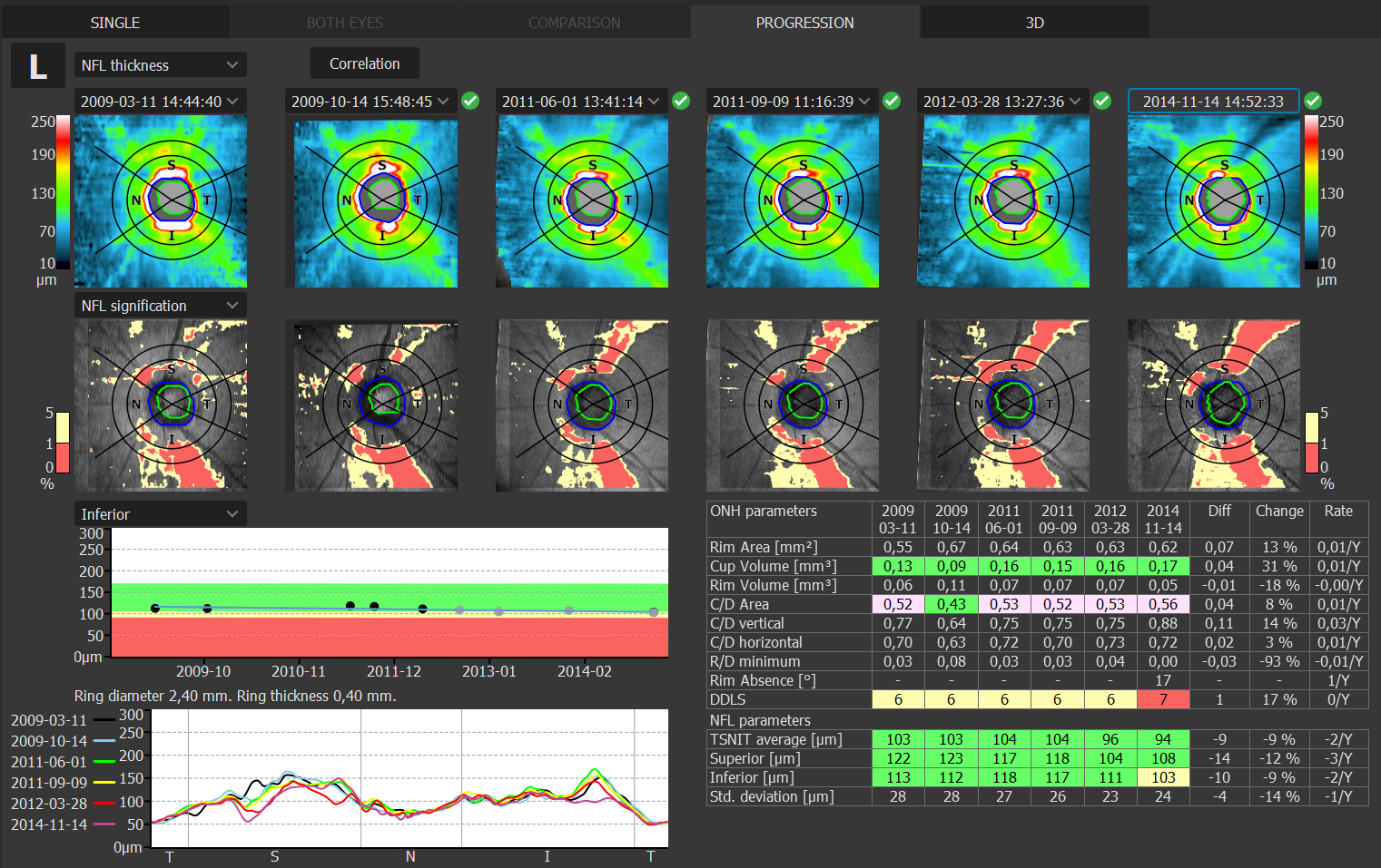
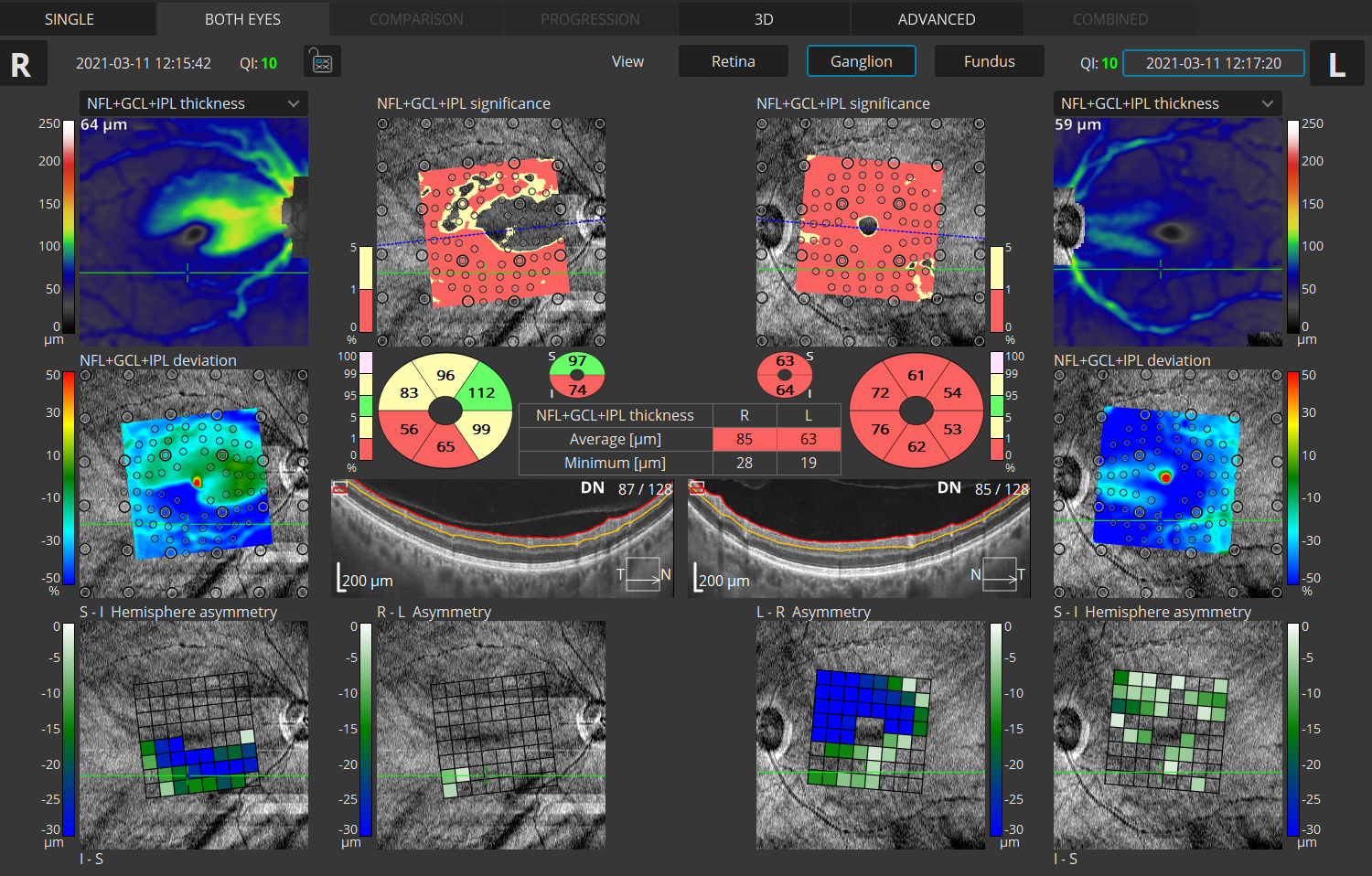
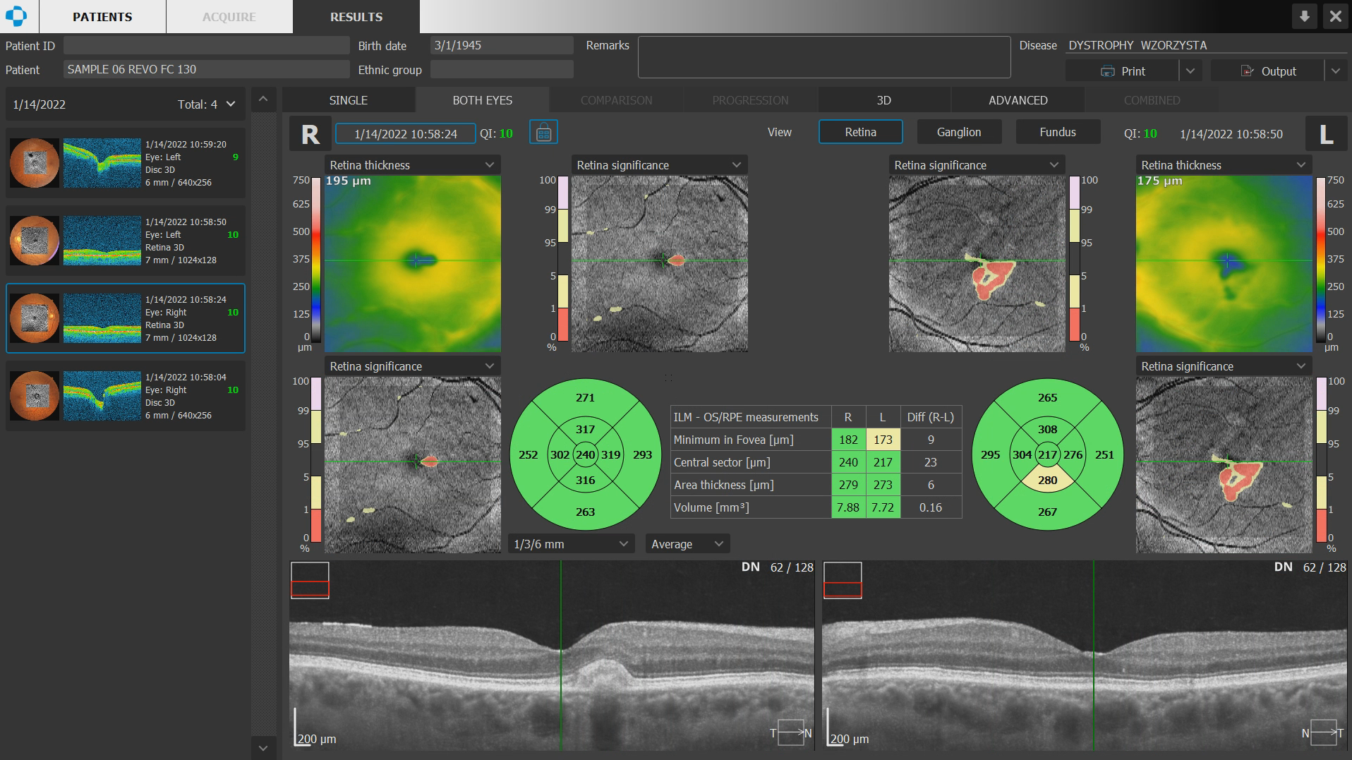
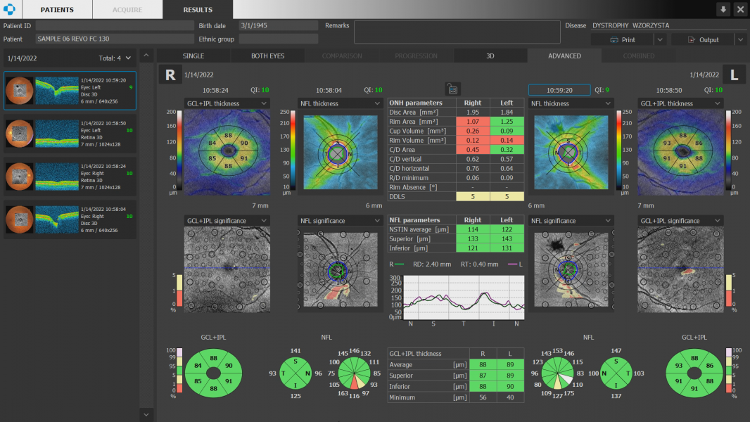
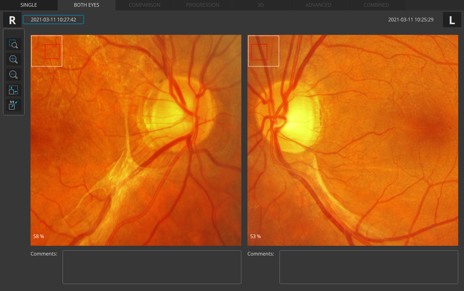
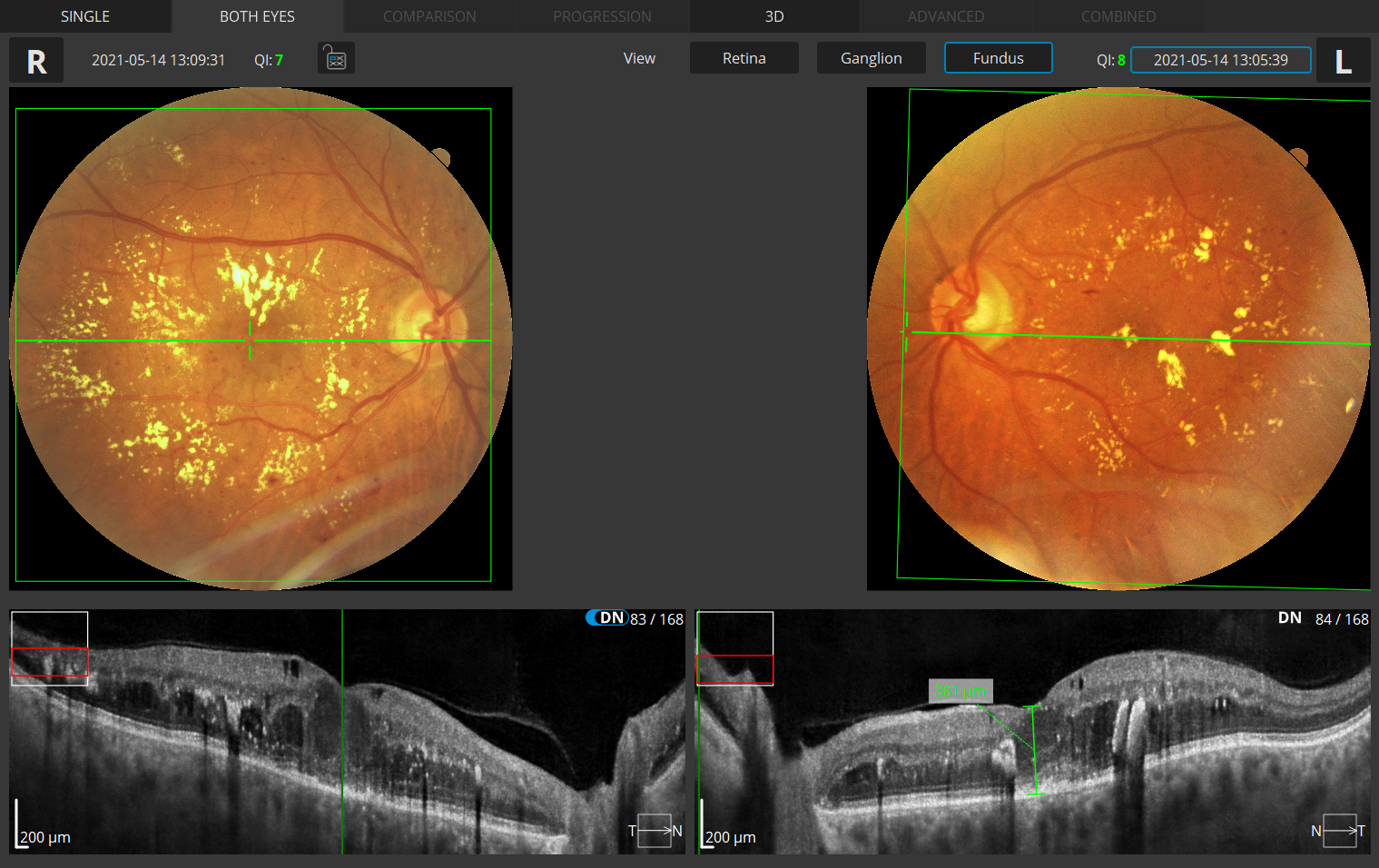
Structure + Function (S+F)
Comprehensive glaucoma solution that combines REVO OCT and PTS Visual Field results. S+F takes the diagnostic approach of the Hood report.
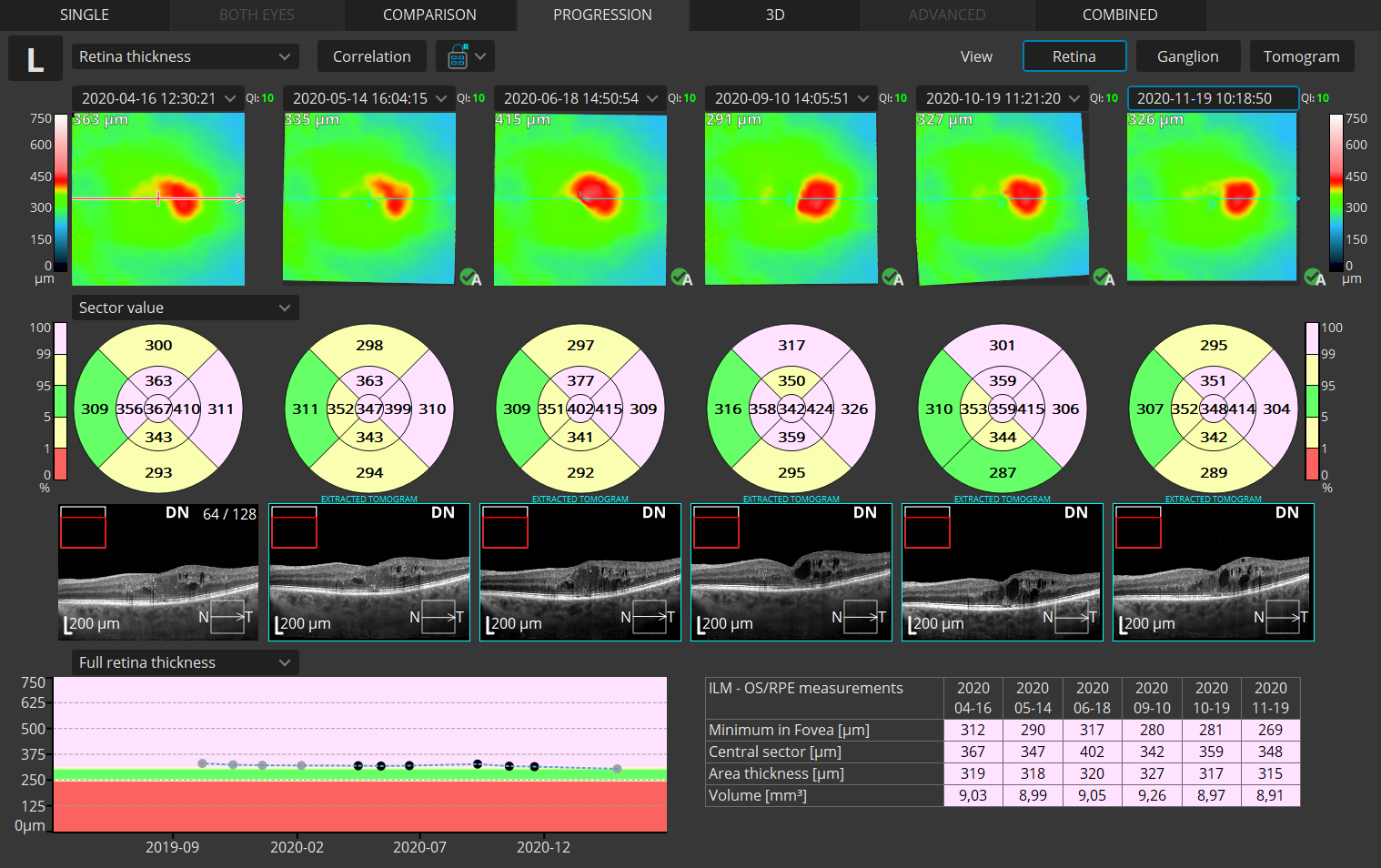
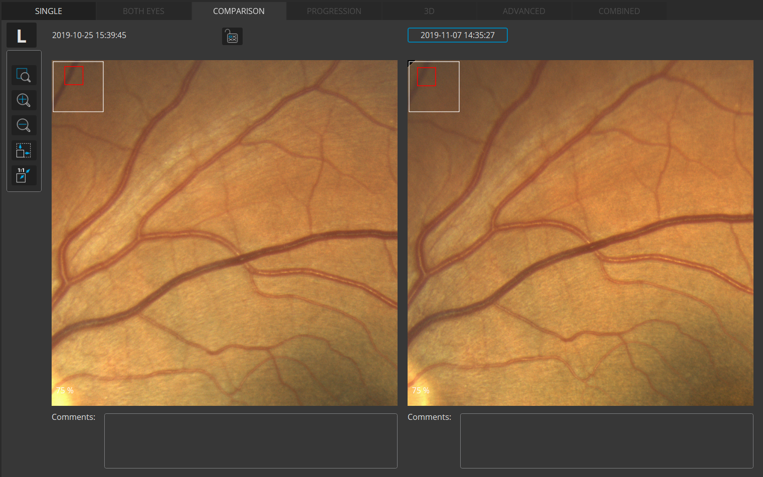
Progression Analysis
Gather baselines and follow-ups to monitor and manage disease progression in posterior and anterior scans.
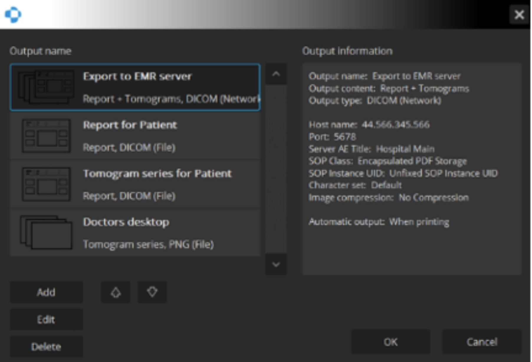
Network Integration
A proficient networking solution with DICOM and EMR capabilities. Quickly and easily export to a desired location.